
Advancements in genetics, cell biology, and medical diagnostics rely on precise laboratory techniques and high-quality imaging technologies. Among the most important tools used in research laboratories today are hypotonic solutions and fluorescence microscopy. These methods help scientists prepare biological samples, visualize cellular structures, and analyze genetic material with exceptional accuracy.
As research continues to uncover the complexities of human health and disease, the combination of proper sample preparation and advanced imaging has become increasingly important. Laboratories worldwide depend on these techniques to generate reliable data that supports scientific discoveries and clinical decision-making.
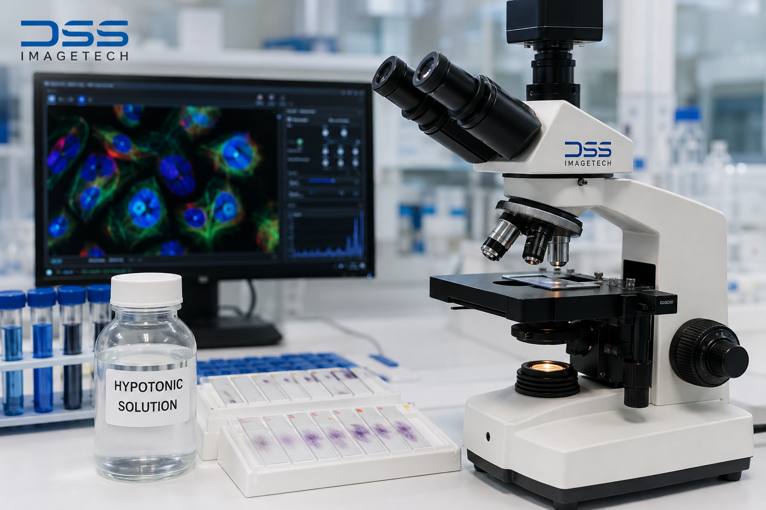
Understanding Hypotonic Solutions
A hypotonic solution contains a lower concentration of dissolved substances compared to the interior of a cell. When cells are exposed to this environment, water enters the cells through osmosis, causing them to expand or swell.
This swelling process is particularly valuable in cytogenetic and chromosome studies. By increasing cell volume, chromosomes become more separated and easier to observe under a microscope. This improved visibility allows researchers to perform detailed genetic analyses and identify abnormalities that may otherwise remain hidden.
In laboratory settings, hypotonic solutions are commonly used during:
- Chromosome preparation
- Karyotyping procedures
- Genetic disorder investigations
- Cancer cytogenetics research
- Prenatal diagnostic testing
Proper sample preparation is a critical step in ensuring accurate laboratory results, making hypotonic solutions an essential component of many biological workflows.
The Science Behind Fluorescence Microscopy
While sample preparation is important, visualization is equally critical. This is where fluorescence microscopy plays a transformative role in modern science.
Unlike traditional light microscopy, fluorescence microscopy uses fluorescent dyes, proteins, or probes that emit light when exposed to specific wavelengths. These fluorescent markers allow researchers to target and visualize specific structures within cells and tissues.
The technique provides high contrast and exceptional specificity, making it possible to study biological processes that are difficult to detect using conventional imaging methods.
Researchers use fluorescence microscopy to:
- Examine cellular structures
- Track protein interactions
- Study DNA and RNA sequences
- Investigate disease mechanisms
- Monitor cellular responses to treatments
Its ability to reveal detailed molecular information has made fluorescence microscopy a cornerstone of biomedical and life science research.
Why These Technologies Work Together
One of the most significant applications of hypotonic solutions and fluorescence microscopy is in chromosome analysis and genetic research.
Before chromosomes can be examined, cells are often treated with hypotonic solutions to encourage swelling and chromosome spreading. This preparation creates optimal conditions for visualization.
Scientists then use fluorescence microscopy along with fluorescent probes to identify specific chromosome regions, genes, or molecular markers. The resulting images provide valuable insights into chromosomal abnormalities, genetic mutations, and disease-related changes.
This combined approach helps researchers achieve greater accuracy and supports the development of more effective diagnostic methods.
Supporting Scientific Excellence with DSS Imagetech
Modern laboratories require reliable imaging solutions to meet the growing demands of research and diagnostics. DSS Imagetech has been a trusted name in the field of microscopy, life sciences, and imaging technologies, supporting researchers, healthcare professionals, and academic institutions across India.
Through its advanced microscopy solutions and scientific imaging systems, DSS Imagetech helps laboratories perform high-precision studies involving fluorescence microscopy, chromosome analysis, cell imaging, and molecular research. The company’s commitment to innovation and quality enables scientists to generate accurate and reproducible results for both research and clinical applications.
By providing access to cutting-edge imaging technologies, DSS Imagetech plays an important role in advancing scientific discoveries and improving laboratory workflows.
Applications in Healthcare and Research
The integration of hypotonic solutions and fluorescence microscopy has contributed significantly to advances in healthcare and scientific research.
Cancer Research
Researchers use these techniques to identify chromosomal changes associated with various cancers. Early detection of genetic abnormalities can support treatment planning and improve patient outcomes.
Genetic Testing
Genetic laboratories rely on chromosome preparation and fluorescence-based imaging to diagnose inherited disorders and evaluate genetic risks.
Drug Discovery
Pharmaceutical researchers use fluorescence microscopy to observe how cells respond to potential treatments, helping accelerate the development of new therapies.
Academic and Clinical Research
Universities, research institutes, and diagnostic laboratories use these methods to investigate cellular behavior and better understand disease mechanisms.
Conclusion
The success of modern biological and medical research depends on both accurate sample preparation and advanced visualization technologies. Hypotonic solutions enable effective chromosome and cell preparation, while fluorescence microscopy provides the detailed imaging needed to study complex biological structures.
Together, these techniques support breakthroughs in genetics, diagnostics, cancer research, and drug development. With innovative imaging providers like DSS Imagetech supporting the scientific community, researchers have access to the tools they need to explore new discoveries and improve healthcare outcomes worldwide.